


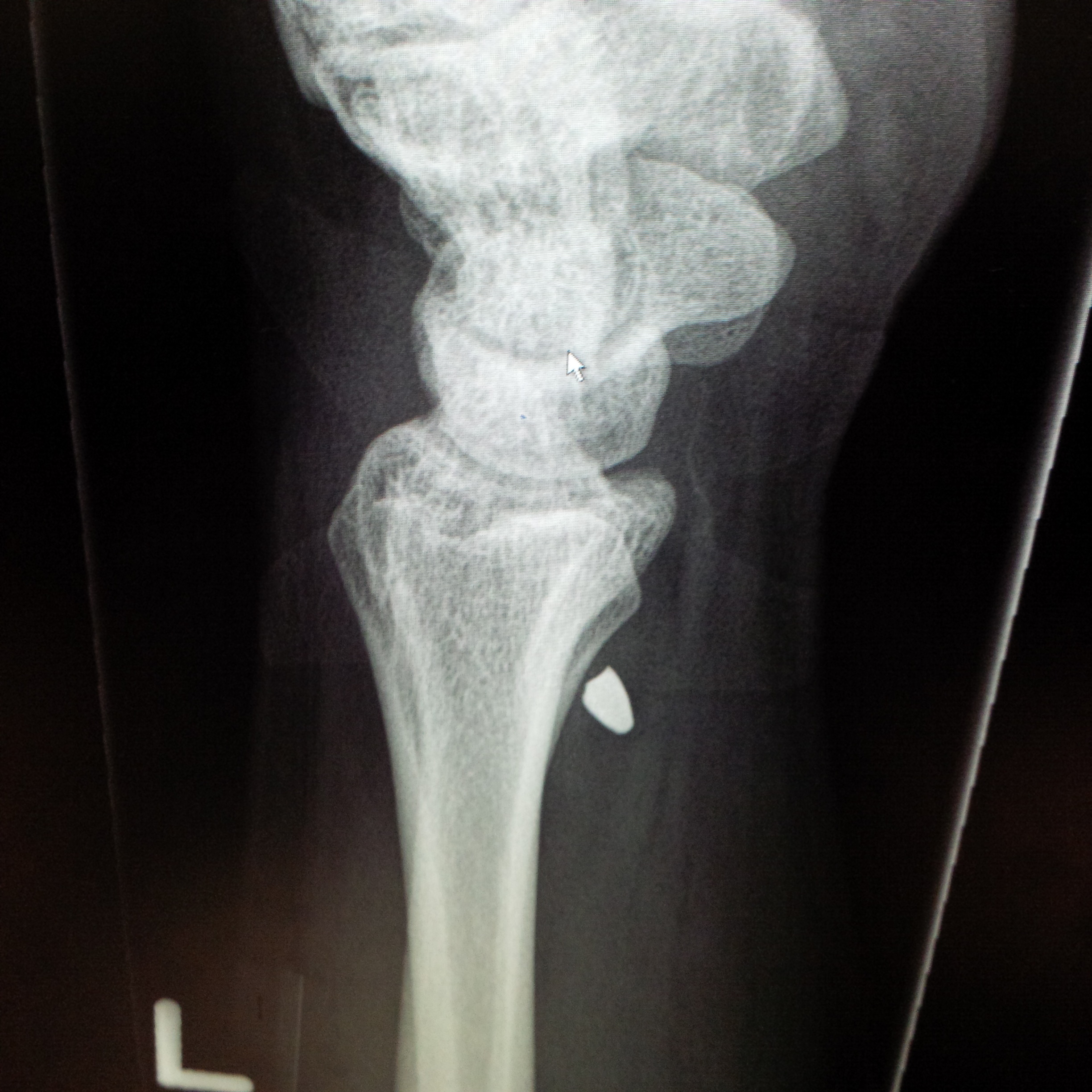
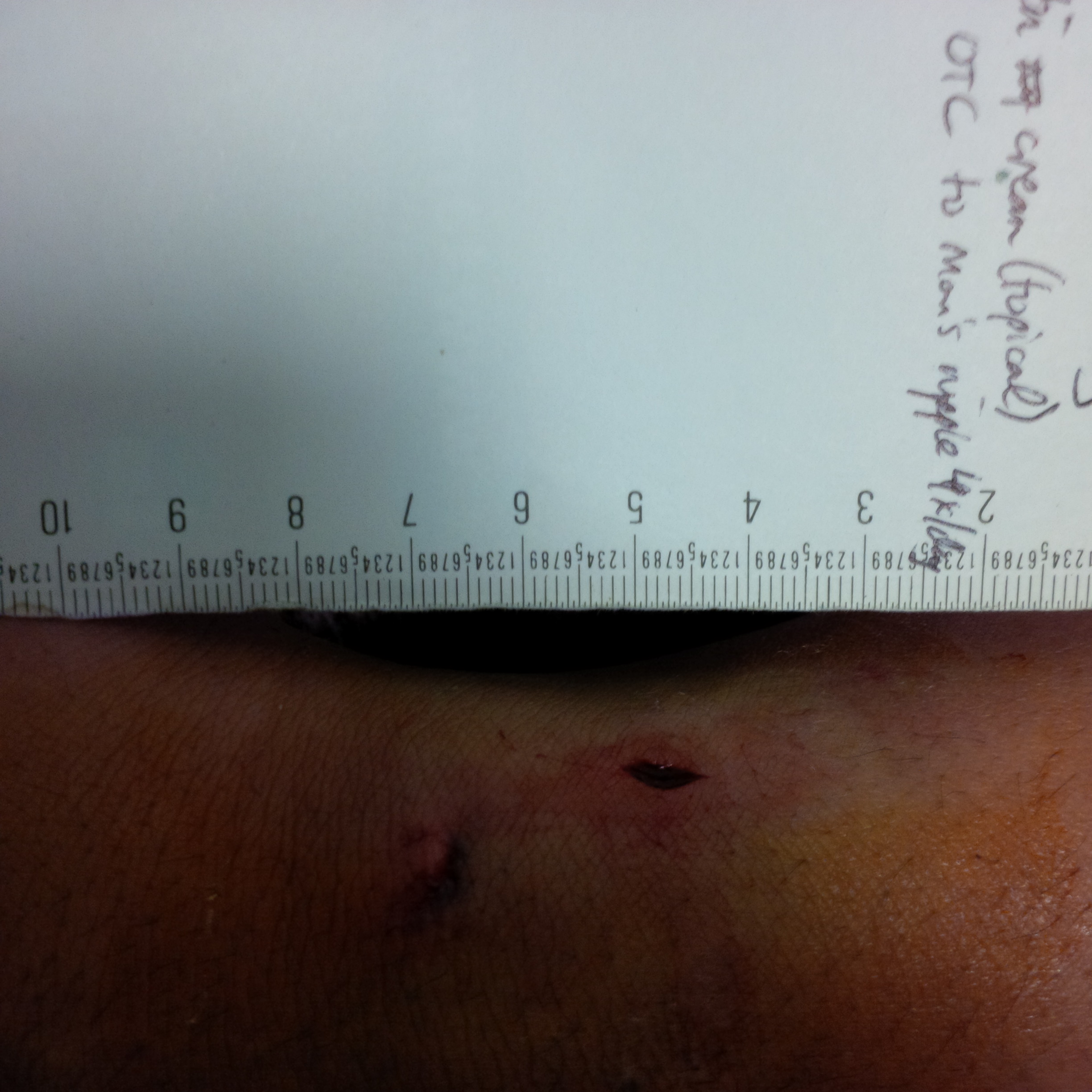
40 year old mechanic was hammering metal when his hammer broke. He immediately felt pain in his wrist. He was concerned part of the hammer was stuck in his wrist. He had an 8 mm laceration to the volar surface of his wrist. He has normal ROM, normal pulses, normal Allen test, and normal sensation. He was sent for an x-ray to look for a metallic foreign body.
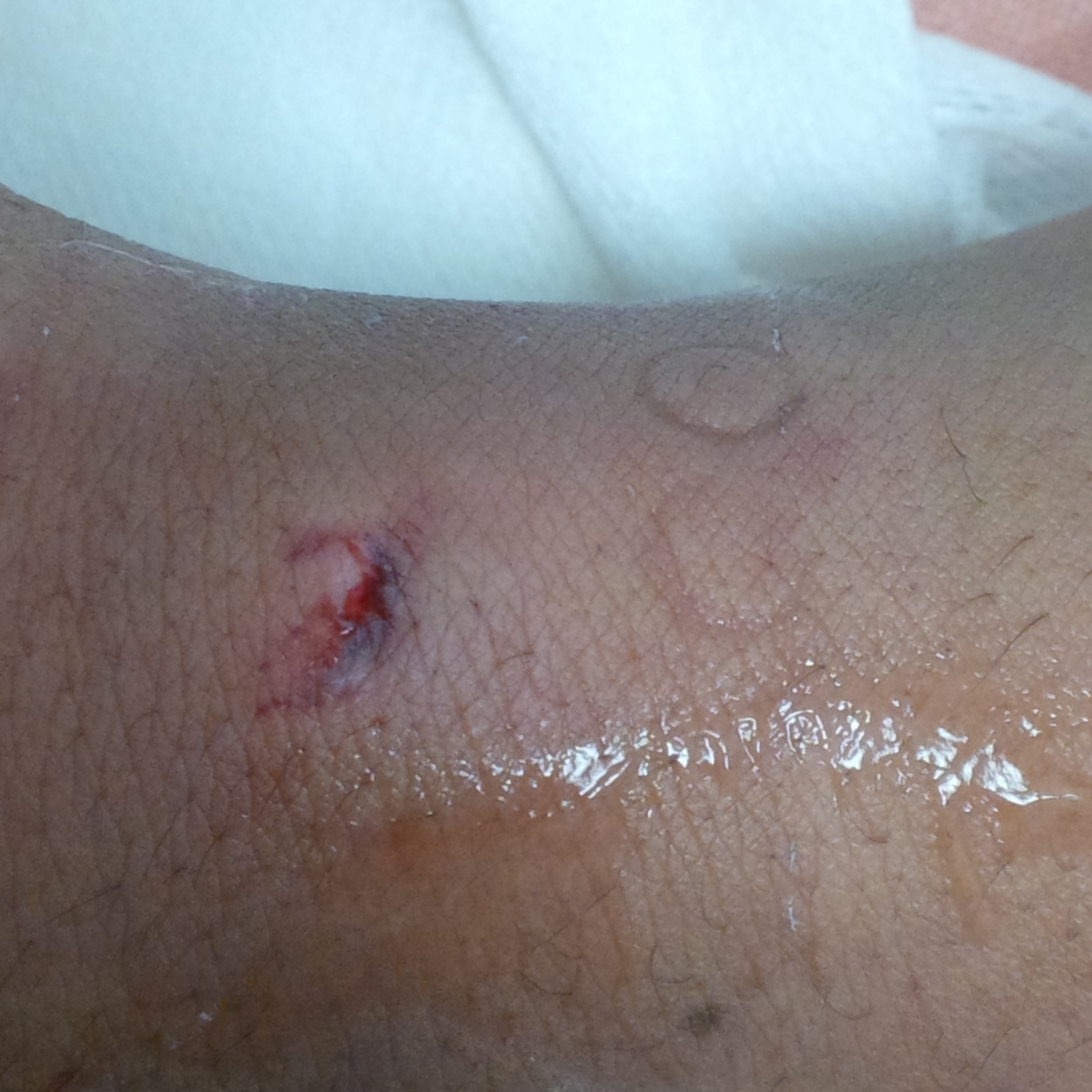

However, on palpating near the wound, no FB is felt.
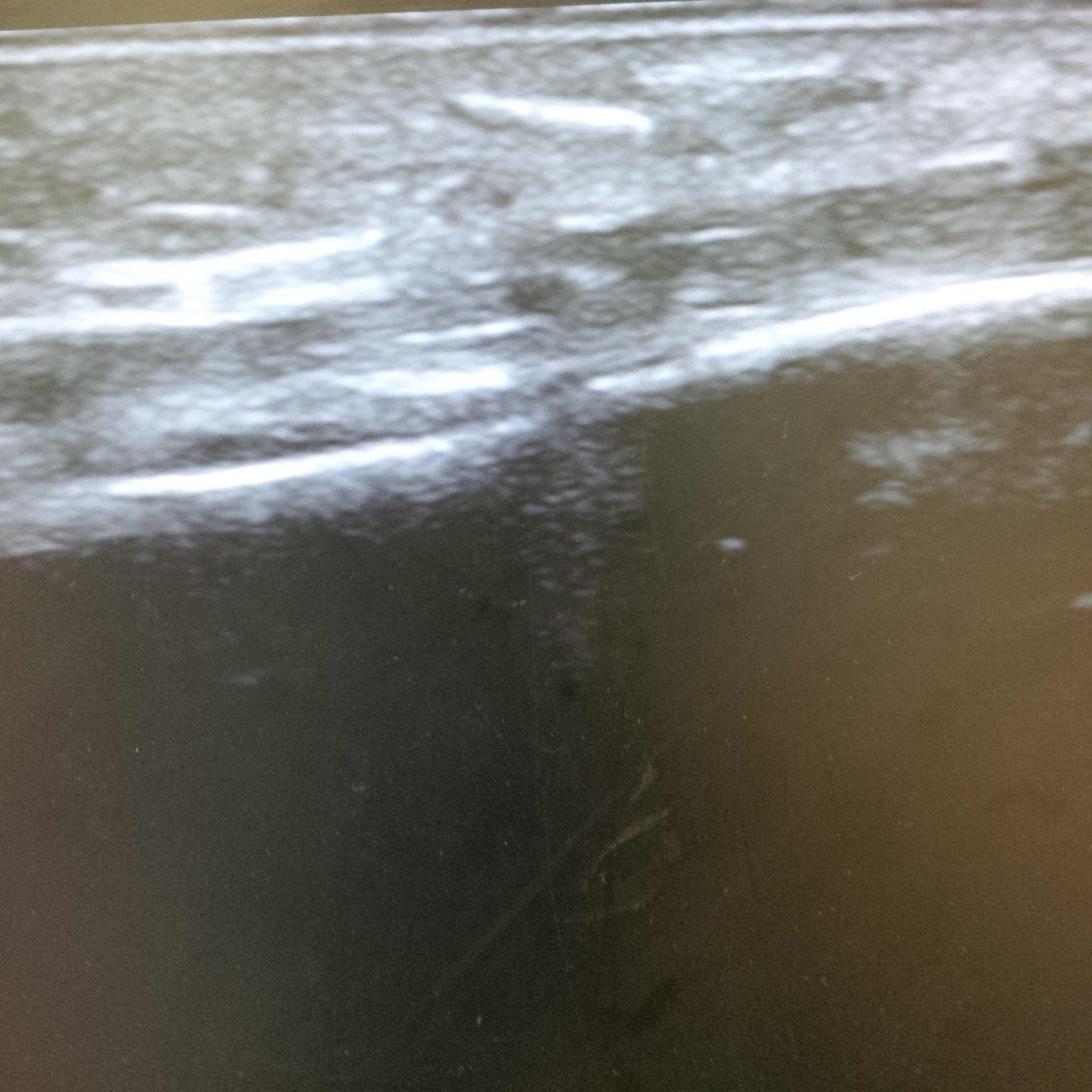
A bedside ultrasound was performed and the metallic foreign body is found. However, the location of the foreign body was not near the location of the laceration.
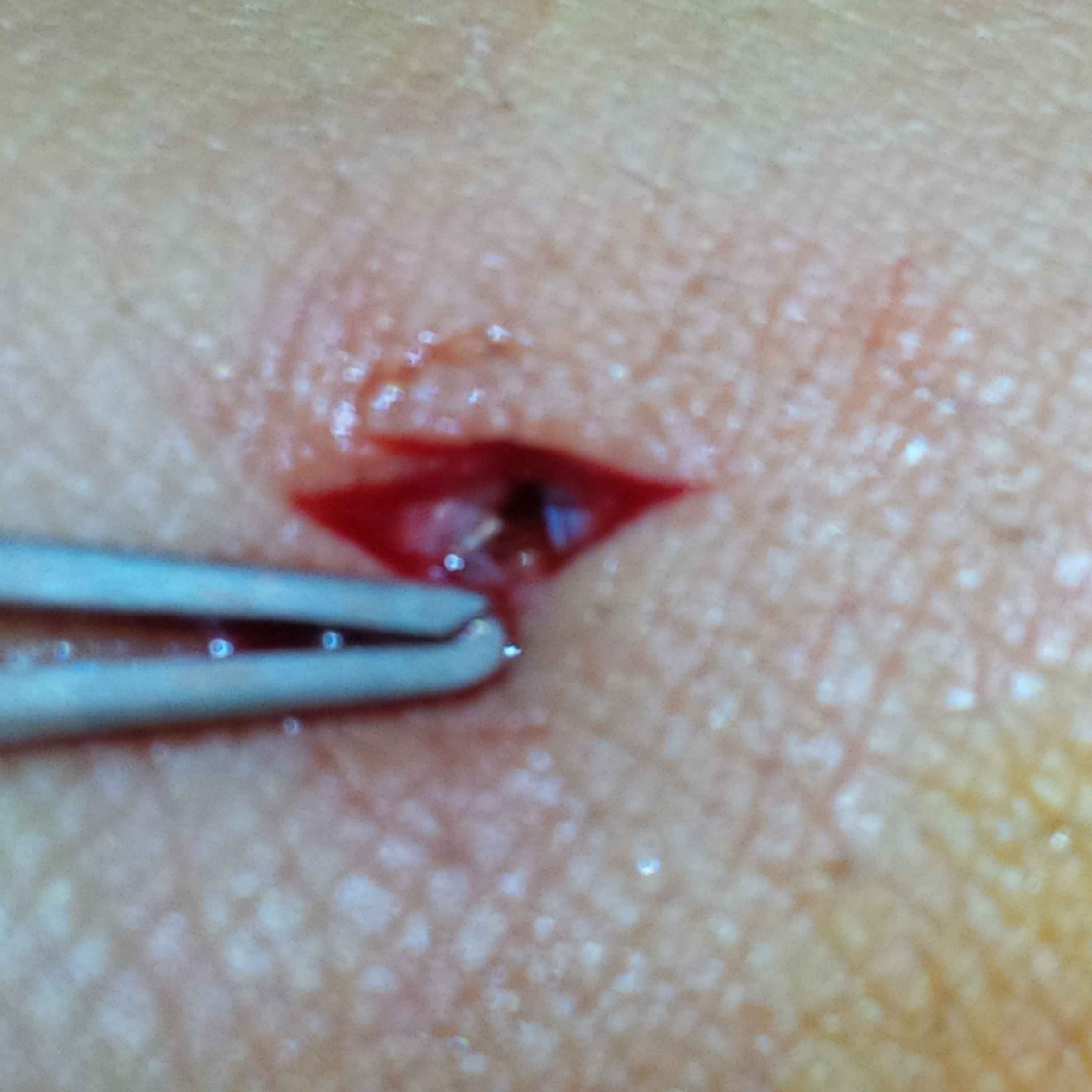
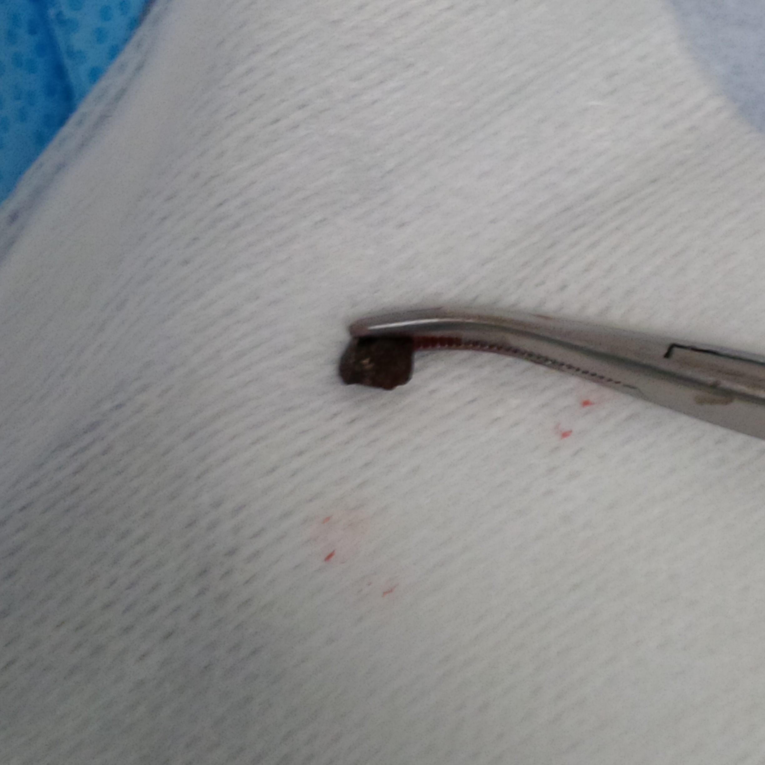
The FB is marked out with the ultrasound probe and a small laceration is made in the skin over the FB. The FB is identified and removed.
Circles to the Right of the wound indicate the location of the FB
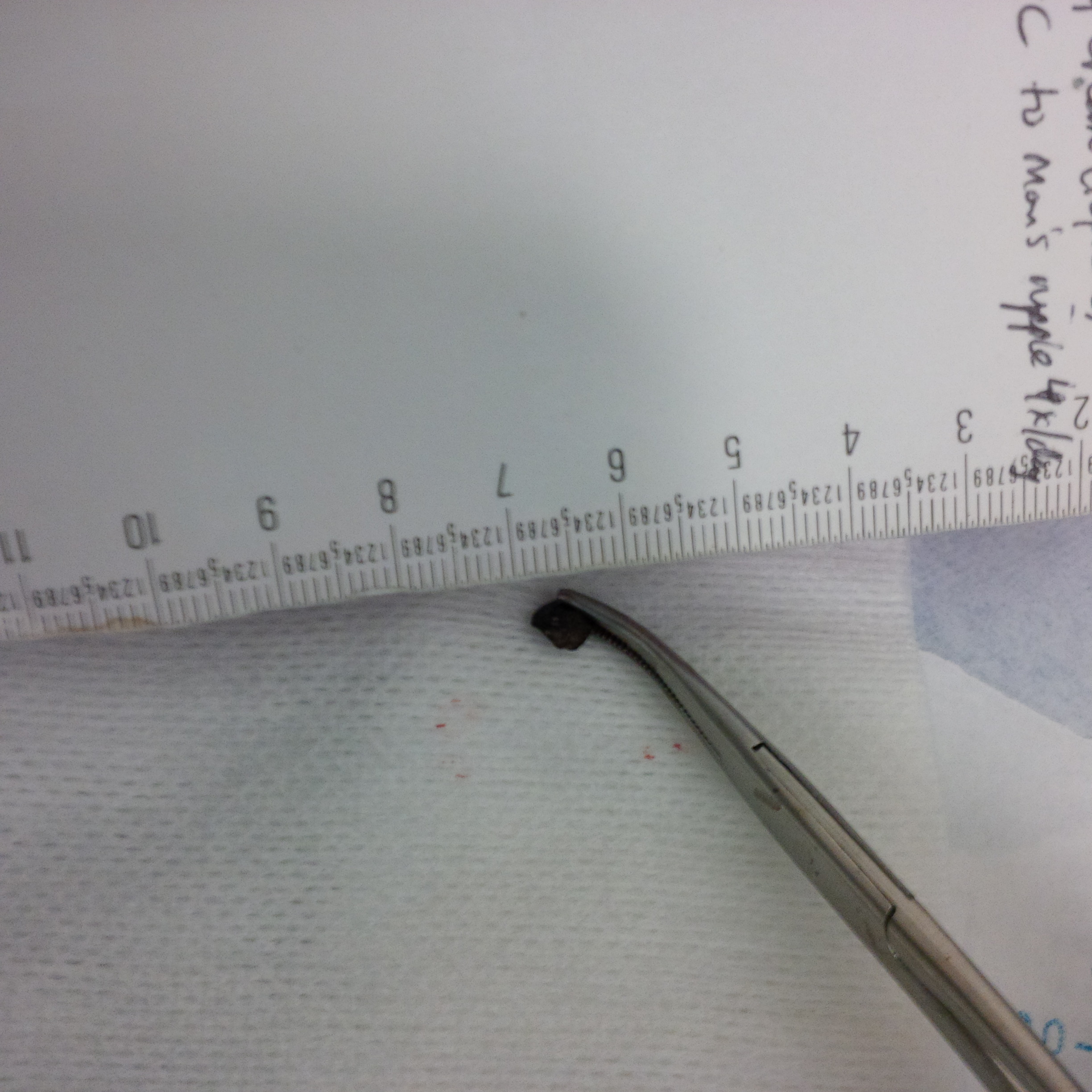
The incision was made 2 cm away from the entrance wound. The FB was 6 mm and the incision made was 7 mm. By using bedside ultrasound, the FB was measured, the location was identified, the skin was landmarked, and the FB was easily retrieved.
Without the ultrasound, the initial entrance would have been extended until the FB was found leaving a very large wound.
Learning Points: using bedside ultrasound can aide not only in finding and removing the FB, but also leads to less invasive probing.
- Foreign Body Removal - November 25, 2014