Case:
A 65-year old healthy lady presents with left shoulder pain after sustaining a fall on outstretched arm injury while walking her dog. She complains about inability to move her left shoulder and severe pain. There was no associated head or neck injury. She has no prior history of shoulder dislocation.
She is hemodynamically stable and you note full distal sensation and motor function in the left extremity. There is an obvious squared off deformity of her left shoulder. Deltoid sensation is in tact.
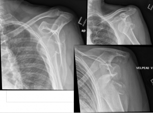
This lady has an obvious left-sided shoulder dislocation and 3-views of the shoulder are ordered.
But in the intrepid ultrasound fellows happen to be lurking in the department… can we use POCUS to diagnose shoulder dislocations?
Background:
The rationale for obtaining a standard set of pre-reduction radiographs are several fold. First, determining whether the humeral head is dislocated anterior or posteriorly has implications on the method of reduction. Second, up to 25% of shoulder dislocations have an associated fracture. This could impact follow-up and degree of immobilization. Third, the standard of care currently mandates radiography (both pre- and post-reduction).
There have been several case reports and series that have investigated using POCUS to diagnose shoulder dislocations. There has been one prospective study investigating the diagnostic performance of POCUS in the ED. “Diagnostic Accuracy of Ultrasonographic Examination in the Management of Shoulder Dislocation in the Emergency Department”Abbasi et al. Ann Emerg Med 2013;62:170-175
Briefly,
- Population: Convenience sample of 73 patients suspected to have shoulder dislocation at 2 academic EDs in Tehran
- Intervention: Shoulder ultrasonography performed by one of two investigators
- Comparison: 3 views on plain radiographs (ant/post; lat; scapular Y)
- Outcome: Predictive characteristics of POCUS for detecting shoulder dislocation and successful reduction
POCUS was found to be 100% sensitive and specific in detecting dislocations. While the sample size was small and there was no mention of the techniques involved, POCUS was able to detect 100% of the associated fractures as well.
Ultrasound Technique
There are three described approaches to using POCUS to diagnose shoulder dislocations.
Which probe? Linear probe is preferred but the curvilinear can be used in patients with larger body habitus
1. Anterior Approach (used in this study) and probably the easiest view to obtain
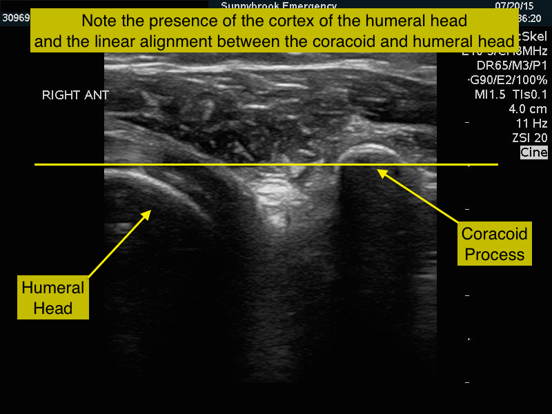
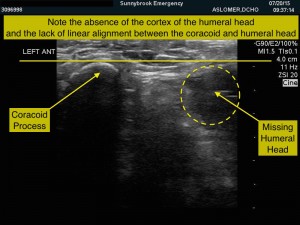
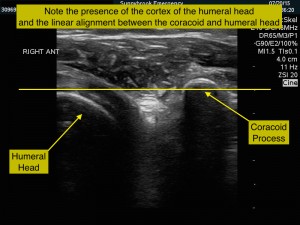
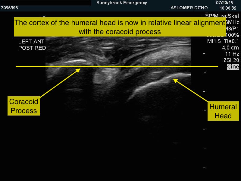
Place the probe on the anterior part of the shoulder on top of the coracoid process in transverse. Focus on the cortices (bright, echogenic, curved). In a normal shoulder, the cortices of the coracoid process and humeral head will be in linear alignment. In a dislocated shoulder, the coracoid process will be visible, but the humeral head will not be in line.
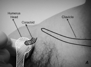
2. Posterior Approach (See the “More Details” Section)
3. Lateral Approach (See the “More Details” Section)
Back to the Patient
Anterior approach with the linear probe yielded these images:
Normal Right Shoulder
Left Shoulder (Abnormal)
Side-by-Side Comparison (abnormal Left; normal Right)
The patient underwent procedural sedation and had her anterior shoulder reduction reduced with no incident. Post reduction ultrasound revealed:
She was placed in a shoulder immobilizer and follow-up was arranged with an orthopedic surgeon for her greater tuberosity fracture.
Clinical Bottom Line
The present study demonstrates excellent sensitivity and specificity in detecting shoulder dislocations (anterior and posterior) in a high-risk population (i.e. those who were suspected to have shoulder pathology based on exam, history, and gestalt). Given how uninvasive and quick the procedure is, it is probably reasonable to use ultrasound to help assess the shoulder joint early in a patient’s clinical course. The exclusive use of POCUS for the management of dislocations without a fracture may be guided by risk factors. Younger people (<40 years old) who present with atraumatic, recurrent dislocations are less likely to have an associated fracture (as described by Emond et al. 2004) and may represent the subgroup of patients who may not need radiography but could be managed with pre- and post-reduction POCUS.
However, at this point, it is probably necessary to get plain radiographs given that there have been no data investigating the diagnostic performance of POCUS for detecting humeral and glenoid fractures. Given the high prevalence of concomitant fractures, the use of POCUS is likely most useful in the context of the post-reduction phase of management. Whilst, the standard of care may not currently support this opinion, the use of POCUS to confirm reduction is likely justified. This is especially true when there is question about whether the joint has been reduced—POCUS could be used to quickly assess the joint even before sedation wears off.
For now, a dislocated shoulder probably does equal two x-rays.
More Details
Lateral approach explored in further detail here:
- How to Use Point-of-Care Ultrasound to Identify Shoulder Dislocation. Christine Riguzzi, MD; Daniel Mantuani, MD; and Arun Nagdev, MD | on February 12, 2014. http://www.acepnow.com/article/use-point-care-ultrasound-identify-shoulder-dislocation/2/
Posterior approach explored in further detail here:
- http://www.emdocs.net/shoulder-ultrasound-reduction-intra-articular-injection/
References
- M. Emond, N. Le Sage, A. Lavoie, et al. Clinical factors predicting fractures associated with an anterior shoulder dislocation. Acad Emerg Med 2004;11:853
- Abbasi et al. Diagnostic Accuracy of Ultrasonographic Examination in the Management of Shoulder Dislocation in the Emergency Department. Ann Emerg Med 2013;62:170-175
- The Meaning of B-Lines - November 15, 2015
- Does shoulder dislocation = 2 x-rays? - August 16, 2015



2 Comments
Great case, thanks!
Anterior shoulder dislocations in pediatric patients: are routine prereduction radiographs necessary?